A Case of Canine Cataracts – Dogster

[ad_1]
Show him, Sadie. See? Look, look! See how she can’t see her cookie?” I was seated across from an 11-year-old snowy Schnauzer that was apparently oblivious to the treat dangling in front of her. I’d known Sadie for six years, since her family fled the frigid winters of upstate New York for the milder climate of coastal Carolina. I’d also seen this coming for a couple of those years (pun intended).
“No matter what I show her, she can’t see it anymore. She constantly bumps into things. And her eyes are so cloudy. Can Sadie see?”
While the answer may have been obvious, delivering the diagnosis to Sadie’s mom required a delicate touch.
“I believe you’re right. I’m afraid her cataracts have finally progressed to the point of severely limiting her vision. She may still see shadows, but I think it’s time we discuss cataract surgery again.”
What Are Cataracts?
Cataracts are one of the most common causes of decreased vision or blindness in dogs. The most frequent reason dogs develop cataracts is genetic. Despite our best efforts, dogs born with a genetic predisposition will likely develop cataracts if they live long enough. In these cases, many dogs develop early signs of cataracts in their late middle- to early-senior years (5 to 12). Other causes include diabetes, eye injuries or infections or certain nutritional deficiencies in young puppies.
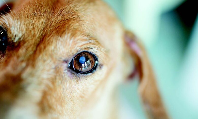
Cataracts are easy to spot due to the cloudiness and whitish opacity they cause framed by the darker iris. In advanced stages, cataracts may look like a crystalline rock inside the eyeball. Sadie was still in the earlier stages, and her eyes had a soft curtain of clouds draping her pupils. The cataracts were now blocking much of the light her retina needed to “see” her surroundings — including the treats her dog parent dangled.
We had recently performed Sadie’s six-month senior blood and urine tests, and no signs of diabetes or other illness was evident. Cataracts usually slowly advance, and based on my history with Sadie, she was due for surgery.
In addition to blindness, a key reason veterinarians encourage surgical removal of a cataract is to prevent further eye damage. Cataracts can luxate or become free-floating within the eye’s chamber, injuring the interior structures, causing severe pain and uveitis. Large or “slipped” cataracts can block the drainage ducts, leading to excruciating glaucoma.
“What about eye drops? Is there any other treatment than surgery?”
She was referring to some misleading websites touting “special cataract-dissolving” eye drops. The primary chemical in question is N-acetylcarnosine (NAC), and, unfortunately, studies have failed to demonstrate success in treating canine cataracts. While we may find NAC has other benefits for our dog’s eyes, treating cataracts isn’t likely to be one of them. While those eye drops can’t make cataracts disappear, there are a few other treatment choices.
Is Surgery Always Necessary?
In dogs with a single, uncomplicated cataract, as long as vision in the normal eye is adequate, surgery may not be needed. In mild cases, anti-inflammatory eye drops, usually a topical NSAID such as diclofenac or ketorolac, combined with a lubricating agent may be used to keep the dog comfortable and reduce the risk of agonizing uveitis. The goal of these medications is to keep the patient comfortable and pain-free. Many dogs can adjust to life with decreased vision or blindness and rely on their sense of smell and hearing to navigate and resume normal activities.
In dogs with bilateral mature or hypermature cataracts, blindness or those experiencing pain or glaucoma, surgical removal of the cataracts is preferred. Before surgery, the veterinary ophthalmologist will conduct tests to make sure the patient’s retina is healthy and able to “see” again after the cataract is extracted. These tests are important because in some instances there may be hidden damage to the retina that would either increase the risk of complications or fail to restore sight after cataract removal.
The majority of canine cases undergo a quick and relatively low-risk procedure called phacoemulsification. This technique involves making a tiny incision in the cornea (the clear front part of the eyeball) followed by inserting a thin, needle-like instrument. The device emits special high-frequency sound waves that dissolve the cataract and suctions the debris away. An artificial lens is then placed where the cataract was and the cornea sutured.
The artificial lens helps improve vision and prevents the world from being reversed and blurry without it. It also reduces the risk of future glaucoma. Studies show that dogs have a 95% vision rate immediately after surgery, and 80% report normal vision for life.
Because Sadie’s cataracts were mature and causing blindness, I referred her to my favorite veterinary ophthalmologist where she underwent surgery the following week. I checked on her within 72 hours post-op, and she literally had a new outlook on life.
“Look, look! See how she follows the treat! Watch her walk around the room without bumping into things! Her eyes are so bright! It’s a miracle, Dr. Ward! We’re so happy!”
As I watched the pair happily parade around the room, I couldn’t help but feel I was witnessing a little miracle. Restoring sight to this elderly dog also restored hope and joy to her family. I jotted “Excellent outcome. Sadie can see.” in her medical record, but that was only a glimpse into her story. On that day, watching Sadie rediscover the glorious world around her while her dog mom rejoiced, she widened my eyes to the sacred bond we share with our dogs.
[ad_2]
Source link


